Diastolic Dysfunction Chart
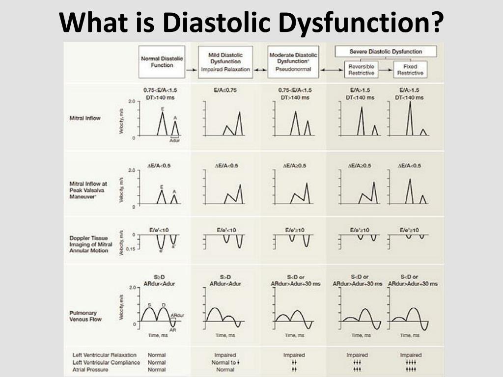
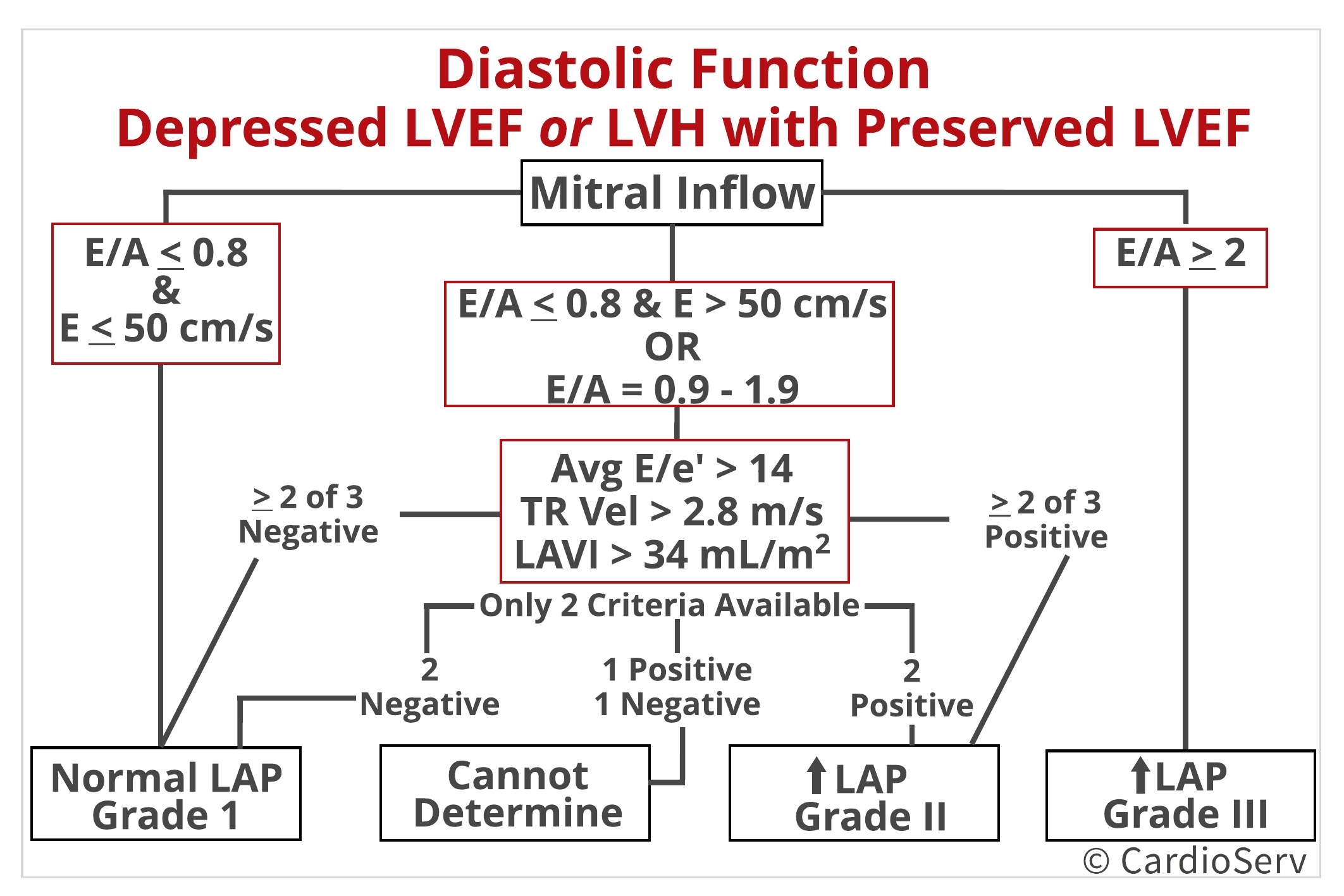
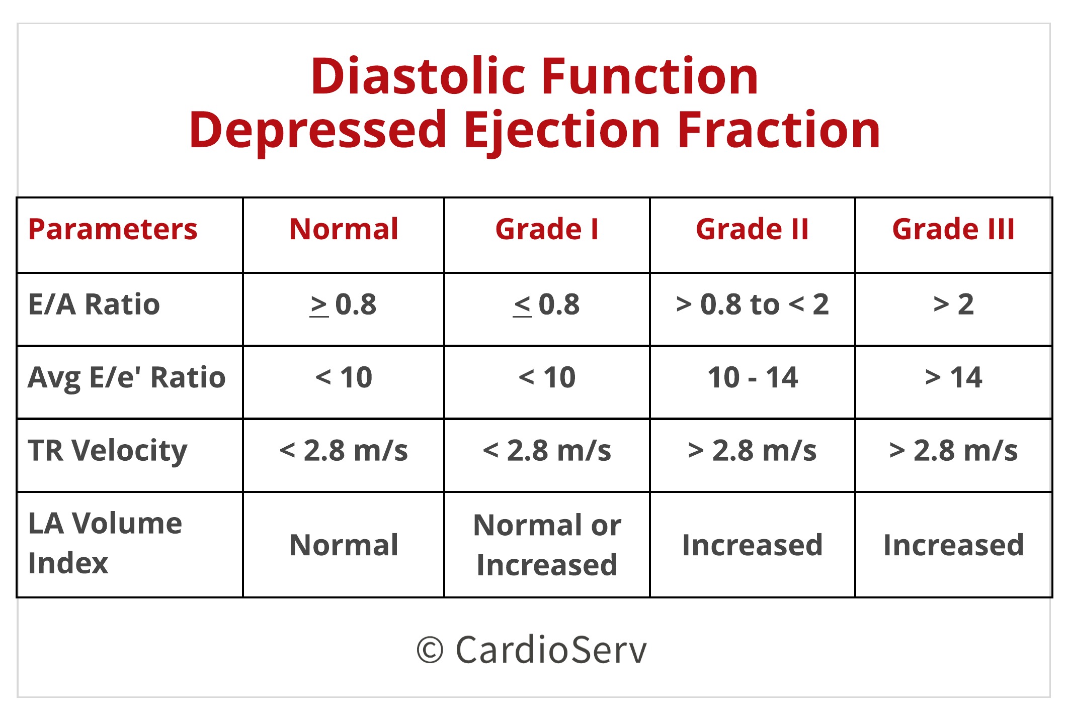
Diastolic Dysfunction Chart - Mitral inflow, tissue doppler, pulmonic vein flow, tricuspid regurgitation velocity, deceleration time, isometric volumetric time, and more. During diastole, your lower heart chambers (ventricles) relax as they fill with blood. Web criteria for diagnosis of lv diastolic dysfunction in patients with normal lvef in jase 2016. Web although diastolic heart failure is clinically and radiographically indistinguishable from systolic heart failure, normal ejection fraction and abnormal diastolic function in the presence of. 1 in contrast, an abnormal filling pattern and progressively greater abnormalities of left filling (impaired relaxation versus pseudonormalized and restricted filling patterns) indicate patients with a progressively increased risk of subsequent mortality. The following section provides recommendations on assessing diastolic function in this group of patients. In pts with normal lvef≥ 50%. Web echocardiographic assessment of lv filling pressures and diastolic dysfunction grade. This disrupts the flow of blood to and from the organs of the body. Web in patients with heart failure and reduced ef (hfref), the main goal is to estimate lv filling pressures and grade the degree of diastolic dysfunction (diastolic dysfunction is presumed to be present in these patients) based on the parameters presented below and the algorithm in figure 8b. This disrupts the flow of blood to and from the organs of the body. Web diastolic function can be estimated from e/a ratio, e’ and deceleration time (dt). Web criteria for diagnosis of lv diastolic dysfunction in patients with normal lvef in jase 2016. In pts with normal lvef≥ 50%. Web in certain clinical situations, conventional echo indices cannot be readily applied to assess diastolic dysfunction. These three methods, as well as several supplementary methods, will now be discussed in detail. Web in patients with heart failure and reduced ef (hfref), the main goal is to estimate lv filling pressures and grade the degree of diastolic dysfunction (diastolic dysfunction is presumed to be present in these patients) based on the parameters presented below and the algorithm in figure 8b. Web echocardiographic assessment of lv filling pressures and diastolic dysfunction grade. During diastole, your lower heart chambers (ventricles) relax as they fill with blood. Web just look at what the american society of echocardiography (ase) guidelines on what you should measure: The following section provides recommendations on assessing diastolic function in this group of patients. Mitral inflow, tissue doppler, pulmonic vein flow, tricuspid regurgitation velocity, deceleration time, isometric volumetric time, and more. Web echocardiographic assessment of lv filling pressures and diastolic dysfunction grade. In pts with normal lvef≥ 50%. Web diastolic dysfunction is when the heart’s ventricles abnormally stiffen, which prevents. During diastole, your lower heart chambers (ventricles) relax as they fill with blood. The following section provides recommendations on assessing diastolic function in this group of patients. Web criteria for diagnosis of lv diastolic dysfunction in patients with normal lvef in jase 2016. In pts with normal lvef≥ 50%. Web in patients with heart failure and reduced ef (hfref), the. Mitral inflow, tissue doppler, pulmonic vein flow, tricuspid regurgitation velocity, deceleration time, isometric volumetric time, and more. Web in patients with heart failure and reduced ef (hfref), the main goal is to estimate lv filling pressures and grade the degree of diastolic dysfunction (diastolic dysfunction is presumed to be present in these patients) based on the parameters presented below and. Blood flow across the mitral valve. Diastolic dysfunction is a problem with diastole, the first part of your heartbeat. These three methods, as well as several supplementary methods, will now be discussed in detail. Web in certain clinical situations, conventional echo indices cannot be readily applied to assess diastolic dysfunction. Diastolic dysfunction may occur when your ventricles are stiff and. These three methods, as well as several supplementary methods, will now be discussed in detail. Web in certain clinical situations, conventional echo indices cannot be readily applied to assess diastolic dysfunction. 1 in contrast, an abnormal filling pattern and progressively greater abnormalities of left filling (impaired relaxation versus pseudonormalized and restricted filling patterns) indicate patients with a progressively increased risk. This disrupts the flow of blood to and from the organs of the body. Web diastolic dysfunction is when the heart’s ventricles abnormally stiffen, which prevents the ventricles from relaxing as they should and prevents them from filling up. Diastolic dysfunction is a problem with diastole, the first part of your heartbeat. These three methods, as well as several supplementary. This disrupts the flow of blood to and from the organs of the body. Web diastolic dysfunction is when the heart’s ventricles abnormally stiffen, which prevents the ventricles from relaxing as they should and prevents them from filling up. In pts with normal lvef≥ 50%. Web in patients with heart failure and reduced ef (hfref), the main goal is to. These three methods, as well as several supplementary methods, will now be discussed in detail. Web criteria for diagnosis of lv diastolic dysfunction in patients with normal lvef in jase 2016. Blood flow across the mitral valve. Web in certain clinical situations, conventional echo indices cannot be readily applied to assess diastolic dysfunction. 1 in contrast, an abnormal filling pattern. Web although diastolic heart failure is clinically and radiographically indistinguishable from systolic heart failure, normal ejection fraction and abnormal diastolic function in the presence of. This disrupts the flow of blood to and from the organs of the body. In pts with normal lvef≥ 50%. Web just look at what the american society of echocardiography (ase) guidelines on what you. Diastolic dysfunction may occur when your ventricles are stiff and don’t relax properly. Diastolic dysfunction is a problem with diastole, the first part of your heartbeat. Web just look at what the american society of echocardiography (ase) guidelines on what you should measure: Mitral inflow, tissue doppler, pulmonic vein flow, tricuspid regurgitation velocity, deceleration time, isometric volumetric time, and more.. Diastolic dysfunction may occur when your ventricles are stiff and don’t relax properly. Diastolic dysfunction is a problem with diastole, the first part of your heartbeat. Web just look at what the american society of echocardiography (ase) guidelines on what you should measure: In pts with normal lvef≥ 50%. Web in patients with heart failure and reduced ef (hfref), the main goal is to estimate lv filling pressures and grade the degree of diastolic dysfunction (diastolic dysfunction is presumed to be present in these patients) based on the parameters presented below and the algorithm in figure 8b. During diastole, your lower heart chambers (ventricles) relax as they fill with blood. The following section provides recommendations on assessing diastolic function in this group of patients. Web diastolic dysfunction is when the heart’s ventricles abnormally stiffen, which prevents the ventricles from relaxing as they should and prevents them from filling up. Web in certain clinical situations, conventional echo indices cannot be readily applied to assess diastolic dysfunction. Web diastolic function can be estimated from e/a ratio, e’ and deceleration time (dt). 1 in contrast, an abnormal filling pattern and progressively greater abnormalities of left filling (impaired relaxation versus pseudonormalized and restricted filling patterns) indicate patients with a progressively increased risk of subsequent mortality. Blood flow across the mitral valve. Mitral inflow, tissue doppler, pulmonic vein flow, tricuspid regurgitation velocity, deceleration time, isometric volumetric time, and more. These three methods, as well as several supplementary methods, will now be discussed in detail.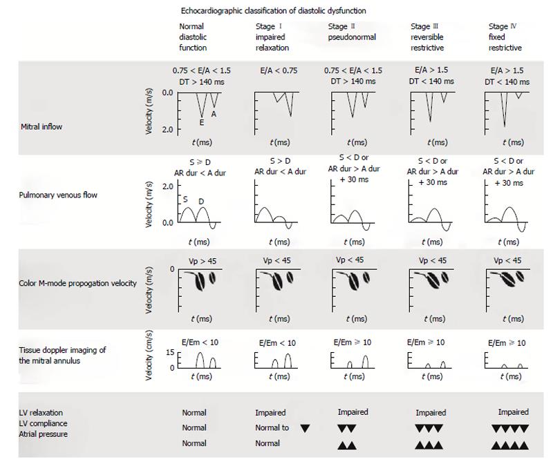
Practical diastology
PPT Diastolic Dysfunction as Diagnosed and Quantified by

diastolic dysfunction Dr.S.Venkatesan MD

How to Measure and Grade Diastolic Dysfunction using Echocardiography

Diastolic Dysfunction Flow Chart
Mastering Diastology Part 2 Cardioserv
Mastering Diastology Part 2 Cardioserv

Alfa img Showing > Diastolic Dysfunction Chart

Diastolic dysfunction Learn the Heart

Lv Diastolic Dysfunction Grade 1 IUCN Water
This Disrupts The Flow Of Blood To And From The Organs Of The Body.
Web Echocardiographic Assessment Of Lv Filling Pressures And Diastolic Dysfunction Grade.
Web Criteria For Diagnosis Of Lv Diastolic Dysfunction In Patients With Normal Lvef In Jase 2016.
Web Although Diastolic Heart Failure Is Clinically And Radiographically Indistinguishable From Systolic Heart Failure, Normal Ejection Fraction And Abnormal Diastolic Function In The Presence Of.
Related Post: